Cell丨靶向PRTG显示对三型髓母细胞瘤的潜在疗效
撰文丨我的闺蜜老红帽
髓母细胞瘤是儿童期最常见的恶性脑肿瘤,多发生于人类胚胎期后脑。分子分层描述了髓母细胞瘤的四个转录亚群:SHH、WNT、三型 (Gr3-MB) 和四型 (Gr4-MB)。Gr3-MB与低生存率和高转移率相关。但是,Gr3-MB肿瘤发生的细胞和分子机制尚不清楚。
人类大脑的发育,包括菱脑唇 (rhombic lip,简称RL) 的空间区隔,与其他哺乳动物明显不同。在人类在后脑神经板和随后的RL脑室区 (RL ventricular zone,简称RLVZ) 中,发现妊娠早期的progenin (PRTG) 阳性干细胞样细胞位于Gr3-MB细胞结构的顶端。PRTG是一种已知的膜蛋白,在早期神经管发育过程中可防止早衰分化和细胞凋亡。
近期,来自加拿大The Hospital for Sick Children的 Michael D. Taylor 研究组在 Cell 上发表题为 Early rhombic lip Protogenin+stem cells in a human-specific neurovascular niche initiate and maintain group 3 medulloblastoma 的文章, 发现三型髓母细胞瘤可能是由孕早期RLVZ中低PRTG 阳性干细胞引发的,并且由PRTG 阳性细胞和内皮细胞形成的神经血管生态位所维持。在小鼠模型中,靶向PRTG显示对三型髓母细胞瘤的疗效。
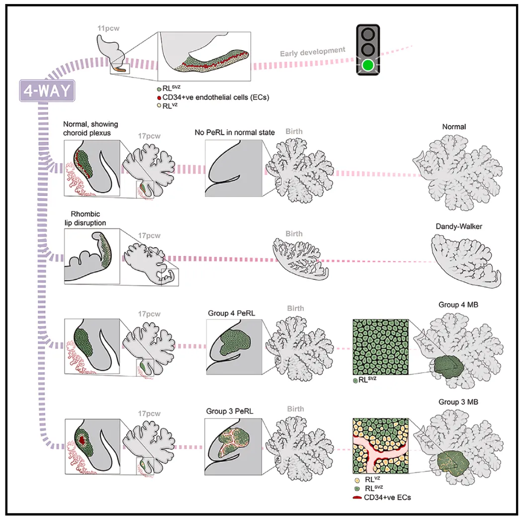
在正常人类RLVZ和Gr3-MB中,PRTG 阳性 细胞生长在CD34 阳性未成熟血管丛附近,这提示了存在维持干细胞的血管周围生态位。RLVZ间插性血管丛 (interposed vascular plexus,简称IVP) 缺损与严重小脑发育不全有关。与之相反的是,作者的研究结果表明,在动物模型中,Gr3-MB顶端干细胞维持了一个持续的未成熟的共生神经血管生态位,其内皮细胞在转录谱上与RLVZ IVP相似。
学界致力于识别癌症干细胞,是基于这一假设:即切除这一小部分癌症干细胞,将对整个肿瘤产生治疗效果。事实上,在临床前模型中,使用抗PRTG阻断抗体、白喉毒素介导的消融或抗PRTG嵌合抗原受体T细胞 (CAR - T细胞) 来靶向PRTG区域,对Gr3-MB具有一定疗效。
为了鉴定处于Gr3-MB结构顶端的细胞,作者对6例Gr3-MB患者肿瘤进行了单细胞RNA测序 (scRNA-seq),并能够辨别出9种不同的转录特征且无样本偏差。通过识别肿瘤细胞中已知的Gr3-MB癌基因,如MYC和OTX2,以及拷贝数畸变的存在,作者观察到三个不同的Gr3-MB细胞亚群,分别标记为Gr3-A、Gr3-B和Gr3-C。Gr3-A中,分化的谷氨酸能神经元表达基因富集,而Gr3-B富集于表达干样标记物的主动循环细胞。
作者在四周大的人胚胎后脑中发现了一群PRTG 阳性MYC高表达 NESTIN低表达的干细胞(PRTG+MYC+NESTIN-),这些干细胞随后定位于RLVZ。早期PRTG阳性干细胞的致瘤性转化引发Gr3-MB样肿瘤。在RLVZ中,PRTG 阳性干细胞生长在人类特异性IVP附近,这种表型在Gr3-MB中重现,但在其他类型的髓母细胞瘤中没有。Gr3-MB与内皮细胞共培养可以促进肿瘤干细胞生长,内皮细胞呈现不成熟表型。在动物体内模型中,使用白喉毒素系统或嵌合抗原受体T细胞靶向Gr3-MB的PRTG阳性区,对肿瘤具有一定疗效。
人类Gr3-MBs可能起源于早期胚胎RLVZ PRTG干细胞,这些干细胞居住在特定的血管周围生态位。靶向PRTG阳性区和/或血管周围生态位很可能是治疗儿童Gr3-MB的一种新方法。
当然,在正常发育和Gr3-MBs中都发现了人类特有的RL特征 (明显的解剖分裂、RL-IVP、相互依赖的神经血管生态位和大量复发的有丝分裂)。但是智人特有的、在小鼠中未见的表型很难用现有的体内模型系统进行研究。事实上,这些人类特有的变化,在多大程度上与人类小脑中大量的谷氨酸能神经元有关,以及其具体作用,目前尚不清楚。更加值得注意的是,这些人类特有的机制很难通过动物实验来验证。
综上所述,作者的工作显示,PRTG+MYC+NESTIN-干细胞可以在未成熟的血管周围生态位中启动并维持Gr3-MB,使用免疫疗法消融这一区域中的细胞很可能又是一项治疗Gr3-MB的新方法。

参考文献:


1. Cavalli, F.M.G., Remke, M., Rampasek, L., Peacock, J., Shih, D.J.H., Luu, B., Garzia, L., Torchia, J., Nor, C., Morrissy, A.S., et al. (2017). Intertumoral Heterogeneity within Medulloblastoma Subgroups. Cancer Cell 31, 737– 754.e6. https://doi.org/10.1016/j.ccell.2017.05.005.

2. Haldipur, P., Aldinger, K.A., Bernardo, S., Deng, M., Timms, A.E., Over- man, L.M., Winter, C., Lisgo, S.N., Silvestri, E., Razavi, F., et al. (2019). Spatiotemporal expansion of primary progenitor zones in the developing human cerebellum. Science 366, 454–460. https://doi.org/10.1126/sci- ence.aax7526.

3. Aldinger, K.A., Thomson, Z., Phelps, I.G., Haldipur, P., Deng, M., Timms, A.E., Hirano, M., Santpere, G., Roco, C., Rosenberg, A.B., et al. (2021). Spatial and cell type transcriptional landscape of human cerebellar devel- opment. Nat. Neurosci. 24, 1163–1175. https://doi.org/10.1038/s41593- 021-00872-y.

4. Wong, Y.H., Lu, A.C., Wang, Y.C., Cheng, H.C., Chang, C., Chen, P.H., Yu, J.Y., and Fann, M.J. (2010). Protogenin defines a transition stage during embryonic neurogenesis and prevents precocious neuronal differentia- tion. J. Neurosci. 30, 4428–4439. https://doi.org/10.1523/JNEUROSCI. 0473-10.2010.

5. Haldipur, P., Bernardo, S., Aldinger, K.A., Sivakumar, T., Millman, J., Sjo- boen, A.H., Dang, D., Dubocanin, D., Deng, M., Timms, A.E., et al. (2021). Evidence of disrupted rhombic lip development in the pathogenesis of Dandy–Walker malformation. Acta Neuropathol. 142, 761–776. https:// doi.org/10.1007/s00401-021-02355-7.

6. Cheng, Y., Liao, S., Xu, G., Hu, J., Guo, D., Du, F., Contreras, A., Cai, K.Q., Peri, S., Wang, Y., et al. (2020). NeuroD1 Dictates Tumor Cell Differentia- tion in Medulloblastoma. Cell Rep. 31, 107782. https://doi.org/10.1016/j. celrep.2020.107782.

7. Fan, X., Masamsetti, V.P., Sun, J.Q., Engholm-Keller, K., Osteil, P., Stud- dert, J., Graham, M.E., Fossat, N., and Tam, P.P. (2021). TWIST1 and chromatin regulatory proteins interact to guide neural crest cell differenti- ation. eLife 10, e62873. https://doi.org/10.7554/eLife.62873.


责编

制作

排版 | 车洁   校对 | uu


▼滑动查看更多▼

原文摘要(Abstract)

We identify a population of Protogenin-positive (PRTG +ve ) MYC high NESTIN low stem cells in the four-week-old human embryonic hindbrain that subsequently localizes to the ventricular zone of the rhombic lip (RL VZ ). Oncogenic transformation of early Prtg +ve rhombic lip stem cells initiates group 3 medulloblastoma (Gr3-MB)-like tumors. PRTG +ve stem cells grow adjacent to a human-specific interposed vascular plexus in the RL VZ , a phenotype that is recapitulated in Gr3-MB but not in other types of medulloblastoma. Co-culture of Gr3-MB with endothelial cells promotes tumor stem cell growth, with the endothelial cells adopting an immature phenotype. Targeting the PRTG high compartment of Gr3-MBin vivo using either the diphtheria toxin system or chimeric antigen receptor T cells constitutes effective therapy. Human Gr3-MBs likely arise from early embryonic RL VZ PRTG +ve stem cells inhabiting a specific perivascular niche. Targeting the PRTG high compartment and/or the perivascular niche represents an approach to treat children with Gr3-MB.

DOI: 10.1016/j.cell.2024.06.011


THE END