肿瘤生物学中的多重蛋白成像
在过去的十年里,组织成像变得更加丰富多彩。实验和分析方法的进步使得对组织样品中的蛋白质标记物进行高倍数成像成为可能。在单细胞或亚细胞分辨率( single-cell or subcellular resolution )下同时常规成像40-50个标记物的能力,为肿瘤生物学研究开辟了新的前景。
细胞表型、相互作用、通信和空间组织已经变得适合于分子水平的分析,并且应用于患者队列已经确定了几种癌症类型中临床相关的细胞和组织特征。
今天来了解下多重蛋白质成像( multiplex protein imaging )方法在研究肿瘤生物学中的应用。
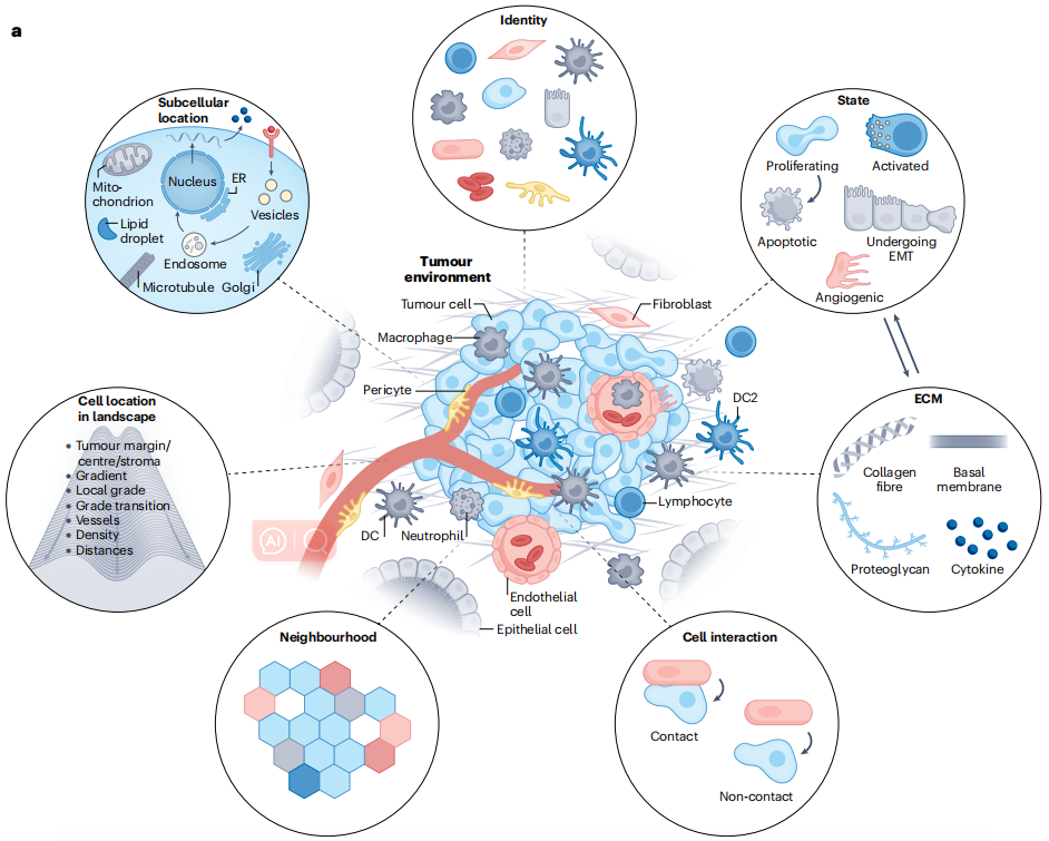
Multiplex protein imaging can interrogate a tumour and its microenvironment across scales
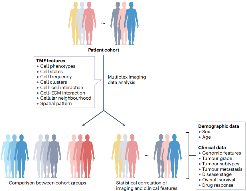
Multiplex protein imaging can be applied to clinical cohorts
An overview of spatial analysis approaches for multiplex protein imaging data
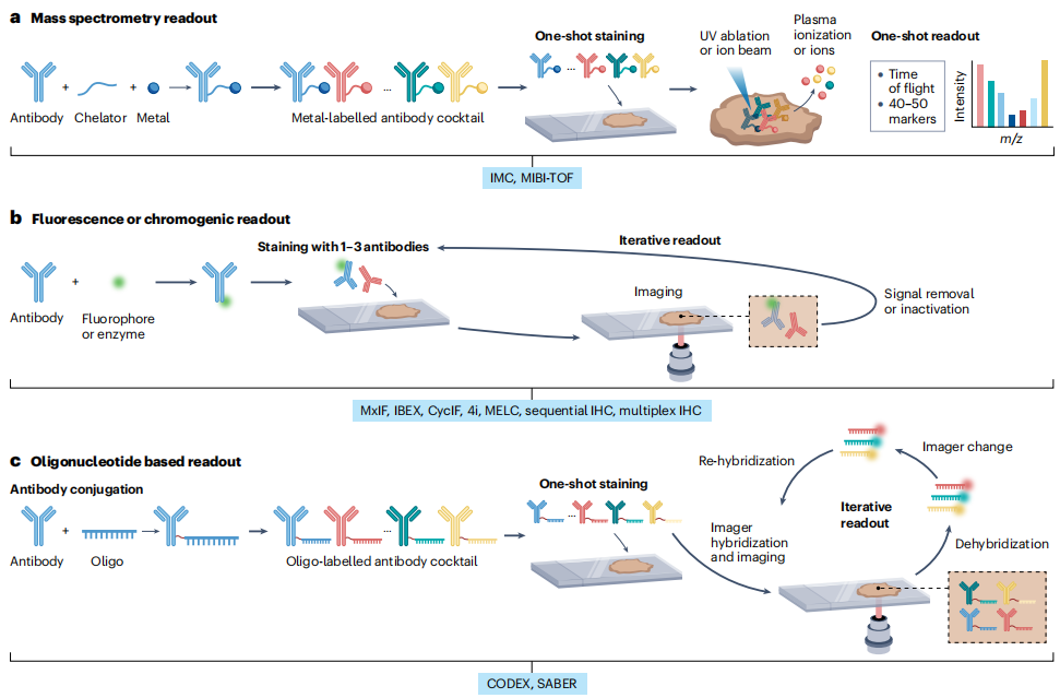
The principles of single-cell multiplex protein imaging methods.
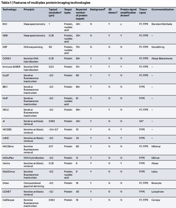
Features of multiplex protein imaging technologies
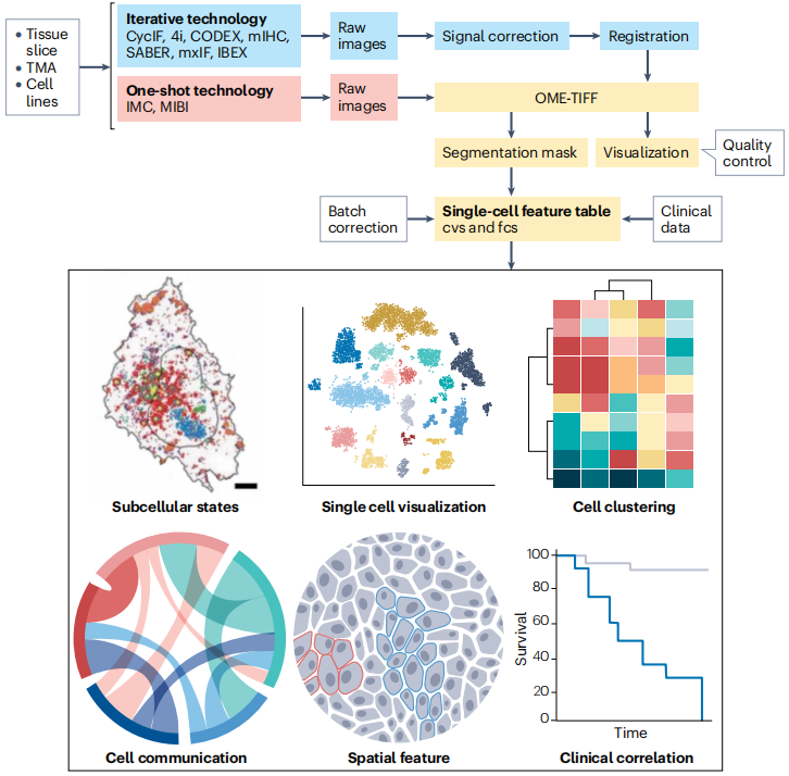
Canonical pipeline for multiplex protein image processing
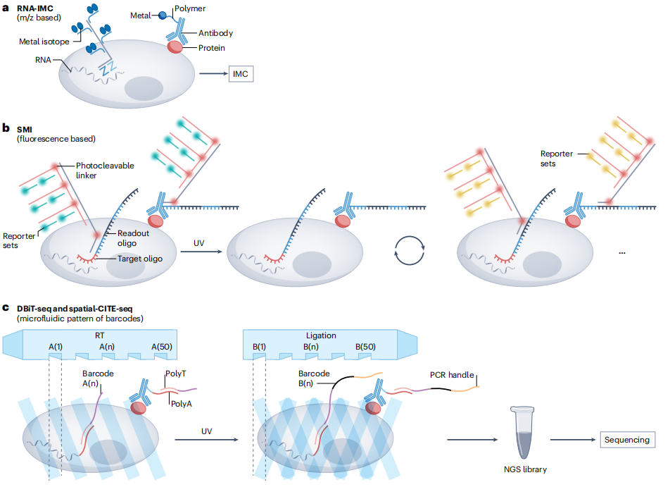
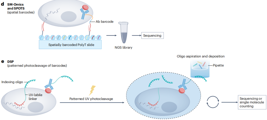
The principles of spatial multi-omics techniques
如果同时包含细胞外和细胞内事件的标记,甚至可以绘制整个信号循环。由于缺乏对肿瘤组织进行单细胞和深层表型分析的可靠方法,到目前为止,特别是在人类样本中,根本不可能探索这些问题。单细胞分辨( Single-cell-resolved )、多蛋白和多模态成像( multimodal imaging )将突破 这一难点。
We envision increasingly colourful and exciting times ahead.
We envision 我们设想, increasingly colourful and exciting 更加丰富多彩和激动
The ability to routinely image 40–50 markers simultaneously, at single-cell or su bcellular resolution, has opened up new vistas in the study of tumour biology.
The ability to .....has opened up new vistas in.....
Here, we review the use of multiplex protein imaging methods to study tumour biology, discuss ongoing attempts to combine these approaches with other forms of spatial omics, and highlight challenges in the feld.
Here, we review ..... , discuss ..... , and highlight .....
参考文献:de Souza, Natalie et al. “Multiplex protein imaging in tumour biology.” Nature reviews. Cancer vol. 24,3 (2024): 171-191. doi:10.1038/s41568-023-00657-4.
版权声明:本文为“乐问号”作者或机构在乐问医学上传并发布,仅代表该作者或机构观点,不代表乐问医学的观点或立场,不能作为个体诊疗依据,如有不适,请结合自身情况寻求医生的针对性治疗。
链接:http://www.lewenyixue.com/2024/04/19/%e8%82%bf%e7%98%a4%e7%94%9f%e7%89%a9%e5%ad%a6%e4%b8%ad%e7%9a%84%e5%a4%9a%e9%87%8d%e8%9b%8b%e7%99%bd%e6%88%90%e5%83%8f/
THE END
